Home » Case Studies » 17th century Umbrian School Madonna and Child
17th century Umbrian School Madonna and Child
Scientific Methods
This is a devotional panel painting depicting the Virgin Mary with the Christ Child, likely dating to the 16th-17th centtury Umbrian school.
Mary is shown half-length, gently inclined toward the Child, her expression soft and introspective. Her lowered eyes and delicate features convey tenderness and humility.
The Christ Child sits on her arm, facing the viewer. He raises one hand in a gesture of blessing, while the other rests naturally by his side.
Both figures wear golden halos, rendered with simple circular forms rather than elaborate punchwork, suggesting a modest workshop or provincial context.
The background is dark and plain, originally intended to heighten the spiritual focus on the figures. Small remnants of decorative elements—such as the gold star on the left—hint at a once richer symbolic setting.
The painting is in poor condition, with extensive paint loss, flaking, abrasions, and areas where the ground layer is exposed. The surface shows widespread cracking and losses, especially in the background and along the edges.
The simplified modeling, warm earth tones, and frontal composition suggest a work intended for private devotion rather than public display.
The overall style bridges Byzantine icon tradition (hieratic pose, gold halos) and Western naturalism (soft facial modeling, childlike proportions).
Even in its damaged state, the painting retains a strong sense of intimacy and devotional purpose, offering insight into everyday religious imagery rather than courtly or monumental art.
CHSOS Collection – item #24
Training: Scientific Methods for Art Examination (December 2025)
This item was analyzed as a case study during the 3-day Training Program on the Scientific Methods for Art Examination hold in December 2025. I would like to thank the participants, Nicole, painting conservator based in Miami and Irwin, radiographer from New York, for conducting the examination and for sharing their expertise while practicing the scientific analysis methods.
Registration open for the Next Training Program!
Technical Photography

The IRF image shows no evidence of cadmium-based pigments.

Infrared Reflectography (IRR)
The Infrared Reflectography (IRR) image was created by stitching together a total of 15 images acquired using our panoramic head VALERIA and an InGaAs camera (320 × 256 pixels). In the IRR region, the blue of the Madonna’s veil and mantle becomes more transparent, allowing additional retouches to be detected. The characteristic behaviour of pigments in the IRR region also aids their identification. For example, the behaviour of this blue pigment—very dark and absorbent in the IR region, but transparent in IRR—is typical of azurite, making it a plausible candidate for this painted area.
X-Radiography
The comparison between the visible image and the radiographic examination (XR) offers valuable insight into both the material condition and the painting process of this 17th-century Umbrian Madonna and Child. In the visible image, the devotional subject is still clearly legible, despite extensive losses of the paint layer and ground, especially in the background and along the contours of the figures. Abrasion and flaking reveal the fragility of the surface and suggest a long history of environmental stress and past interventions.
The radiograph penetrates beyond the pictorial surface, revealing the internal structure of the work. The distribution of denser materials outlines the main figures with greater continuity than is apparent in visible light, confirming the original compositional coherence. Variations in radiopacity suggest differences in pigment composition, particularly in the flesh tones and garments, likely due to the use of lead-based pigments. The radiography also highlights areas of structural damage and previous restorations, providing essential information for conservation decisions and for understanding the artist’s original technique.

Multispectral Imaging (MSI)
We conducted a multispectral imaging examination of the 17th-century Umbrian School “Madonna and Child” as part of our scientific documentation work on this devotional panel painting. Using a simplified protocol with just three band-pass filters centered at 580 nm, 640 nm, and 860 nm, we were able to capture a set of multispectral images that were sufficient to produce a preliminary pigment mapping across the main pictorial areas of the artwork.
These selected wavelengths were chosen to sample key spectral regions where characteristic reflectance differences between historical pigments typically occur, enabling us to distinguish materials with varying spectral behavior without needing a full hyperspectral data cube. The resulting images highlight variations in reflectance associated with different paint components, providing a first approximation of pigment distribution and helping to differentiate between original execution and later retouches.
XRF Spectroscopy
The historical age of the painting is supported by the complex assemblage of elements detected by XRF spectroscopy.
One of the main features of the work is the widespread presence of lead and antimony, suggesting the extensive use of Naples yellow, employed for multiple purposes ranging from the background to the flesh tones and areas associated with gilding. The presence of tin, detected in the gilded areas (point 7) and in the dress (point 5), further suggests the use of lead–tin yellow.
The areas depicting the Madonna and Child (points 3 and 4) show only the elements expected for a painting of this period, mainly lead and iron, indicating that these zones are likely well preserved and free from significant later interventions. Mercury was detected in the red garments (point 5), consistent with the use of vermilion.
The star on the Madonna’s mantle (point 6) contains gold, in addition to the usual presence of lead and antimony, indicating a gilded decoration. This choice suggests a level of material richness that exceeds what might be expected for a modest devotional painting.
Several areas affected by later retouching were identified. In particular, titanium (Ti) was detected in some background areas (points 1 and 2), clearly indicating modern retouches executed with titanium white.
The Madonna’s veil (point 8) shows the presence of cobalt and bismuth, strongly suggesting the original use of smalt. In contrast, the mantle contains copper (point 10), indicating the use of azurite as the blue pigment.
Finally, the analysis of the wooden support revealed the presence of bromine, most likely attributable to brominated biocides or insecticidal treatments applied in the past against woodworm infestation (e.g. methyl bromide fumigation or related preservatives).
Raman Spectroscopy
Raman spectroscopy was used to investigate the materials of the 17th-century Umbrian School Madonna and Child through a non-invasive, in situ examination. The analysis was carried out using our laboratory’s six Raman systems, each operating with a different excitation wavelength, in order to maximize the chances of successful pigment identification while minimizing fluorescence.
Among the available systems, the 830 nm laser proved to be the most effective for this painting. This near-infrared excitation significantly reduced fluorescence effects and allowed the acquisition of clear and interpretable Raman spectra. Using this setup, we successfully identified lead white in the light flesh tones and highlights, as well as vermilion in the red areas of the composition.
Other excitation wavelengths were tested but produced spectra strongly affected by fluorescence or low signal quality. This result highlights the importance of a multi-laser Raman approach in cultural heritage studies, where pigment composition and surface conditions can vary significantly across the artwork.
Reflectance Spectroscopy
Reflectance spectroscopy was carried out on the 17th-century Umbrian School Madonna and Child using our Gorgias Reflectance Spectroscopy system. This non-invasive technique allowed us to investigate the spectral behavior of selected painted areas directly on the surface of the artwork.
The reflectance spectrum acquired from the red vest of the Child Jesus shows a clear inflection point around 580 nm. At first glance, this value differs slightly from the expected inflection point of vermilion, which is typically observed at approximately 600 nm. However, complementary Raman and XRF analyses unequivocally confirmed the presence of vermilion as the main red pigment used in this area.
The observed shift of the inflection point from 600 nm to 580 nm is not unusual in historical paintings. It can be primarily explained by the presence of a red lake glaze applied over the vermilion, which modifies the overall reflectance response. Additional contributing factors include the presence of aged and dirt-embedded varnish layers, pigment mixtures, and complex paint stratigraphy.
This case highlights the importance of interpreting reflectance spectroscopy data within a multi-analytical framework, where spectral shifts are understood as the result of material interactions rather than isolated pigment signatures.
USB microscope
The star decorating the Madonna’s dress was examined using a USB microscope to better characterize its surface and material condition. As expected, the microscopic images reveal surviving traces of the original gilding, confirming that the star was originally executed using a metallic layer rather than painted imitation. Although much of the gilding is now worn or abraded, small areas of reflective material are still visible, particularly along protected edges and recessed zones.
These observations are consistent with the analytical results previously obtained through XRF spectroscopy in the same area. The XRF spectra provided clear indications of the presence of gold, supporting the visual evidence observed under magnification. The combined use of microscopic examination and elemental analysis allows for a more reliable interpretation of the original decorative technique and its current state of preservation.
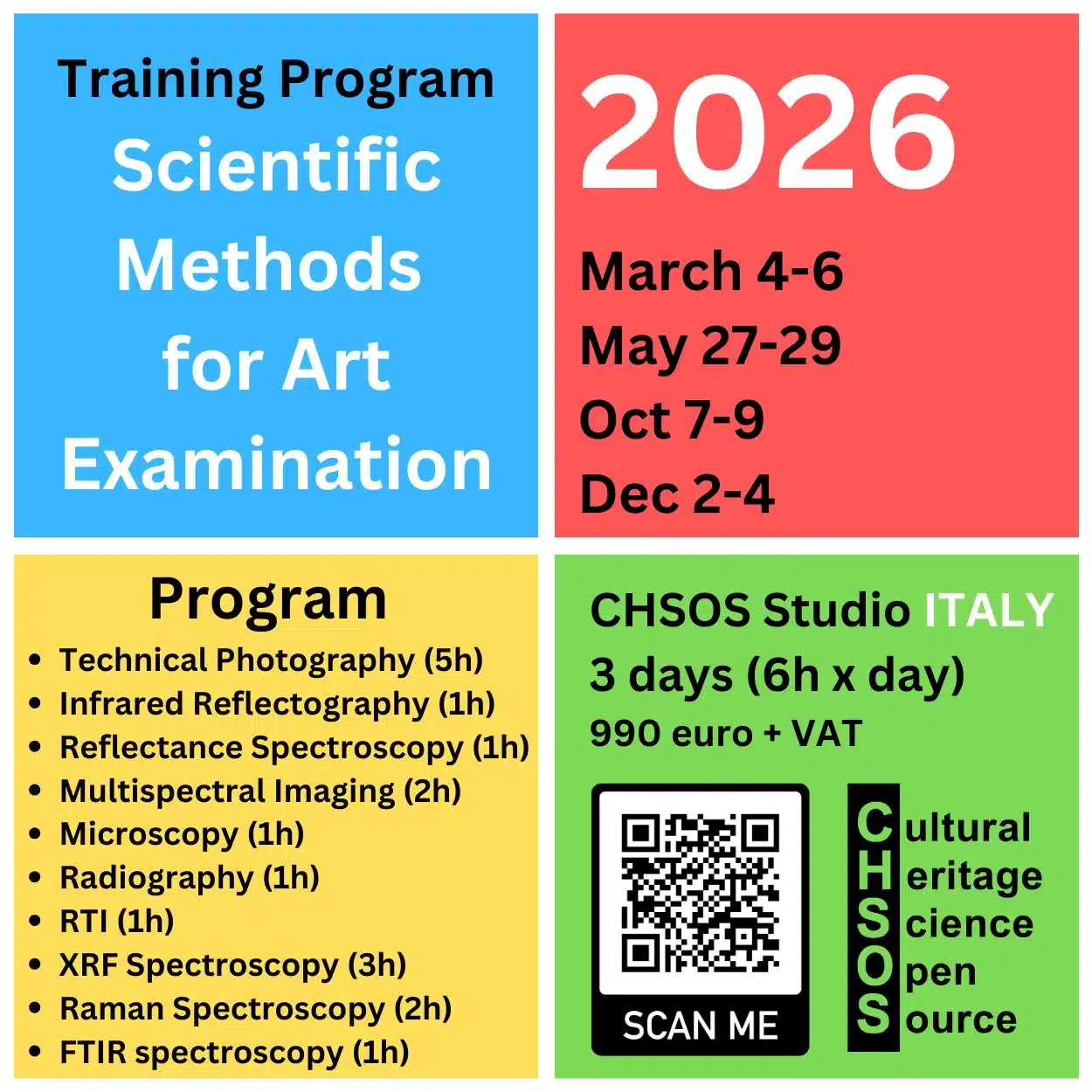
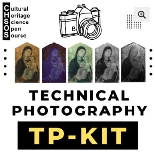
Learn Technical Photography for Art Examination
Technical Photography is one of the most powerful—and often overlooked—tools for the scientific examination of art and archaeology. If you are a conservator, scientist, or art collector and you are not yet familiar with this method, it is truly a missed opportunity. Using simple, affordable equipment and a clear methodology, Technical Photography allows you to reveal underdrawings, retouchings, material differences, and conservation issues in a completely non-invasive way. Far from being complex or inaccessible, it is an easy entry point into scientific analysis. In many cases, Technical Photography represents the first essential step toward a deeper understanding of artworks and archaeological objects.
Scientific Art Examination – Resources:
Getty Conservation Institute (GCI) – USA
The British Museum – Scientific Research Department – UK
Scientific Research Department – The Metropolitan Museum of Art, New York, USA
C2RMF (Centre de Recherche et de Restauration des Musées de France) – France
Rijksmuseum – Science Department – Netherlands


