In this class, we discuss Raman spectroscopy for the identification of iron gall ink, focusing on its application to the two main historical writing supports: parchment and paper.
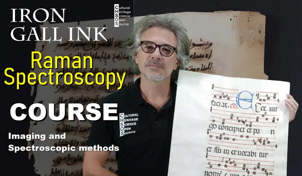
As an example of parchment, we examine a 16th-century antiphonary, where the musical notation is written using iron gall ink. For paper, we analyze a small historical document dated 1527, identifiable from the Roman numerals MDXXV and additional lines in the text. This document was written in Parma, Italy, and provides a representative case study for paper-based supports.
Welcome to our Raman laboratory. This is the space where we perform spectroscopic analyses, including on-site examinations. The mobile bench allows us to bring Raman spectrometers directly to the object under study. For example, we can place the Pigments Checker under the spectrometer and acquire reference spectra directly.
We use several Raman systems with different laser excitation wavelengths: 532 nm (green), 632 nm (red), 785 nm, 830 nm, and 1064 nm (near infrared). The choice of wavelength is crucial because fluorescence, which acts as noise, can obscure the Raman signal. Moving toward longer wavelengths reduces fluorescence, although the Raman signal intensity also decreases. Each application therefore requires a compromise depending on the material, binder, and support.
For iron gall ink on paper and parchment, the 830 nm laser provides the best balance between signal intensity and fluorescence suppression in our setup.
We begin by acquiring a reference Raman spectrum of iron gall ink from the Pigments Checker. This spectrum shows weak Raman features superimposed on strong fluorescence, with characteristic peaks that can be used for comparison.
We then measure the parchment alone, the iron gall ink on parchment, the paper alone, and the iron gall ink on paper. Measuring the supports independently is essential to distinguish which spectral features belong to the substrate and which belong to the ink.
After baseline correction, the spectra are compared. Iron gall ink is identified by two main characteristic Raman peaks, also visible in both parchment and paper samples. The parchment spectrum is dominated by collagen features, including amide bands and skeletal vibrations, while the paper spectrum shows strong cellulose-related peaks.
In conclusion, this lesson demonstrates what to expect when using Raman spectroscopy to identify iron gall ink on parchment and paper, highlighting both the possibilities and limitations of the technique.