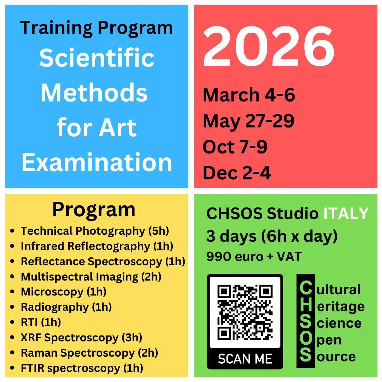
Objectives
- Understand the role of sulfur in pigments.
- Identify the sulfur K-alpha and K-beta lines and their corresponding energy levels.
- Analyze and interpret XRF spectra of sulfur-containing materials, including sulfur minerals, vermilion, gypsum, and ultramarine.
Materials
- Collection of materials for testing:
- Sulfur mineral sample.
- Vermilion (Pigments Checker).
- Gypsum (Pigments Checker).
- Artificial ultramarine (Pigments Checker).
Lesson Plan 1. Introduction to Sulfur in Pigments
- Discuss the importance of sulfur in pigments analysis and its presence in ultramarine, vermilion, and gypsum.
- Use the transmission calculator to demonstrate the feasibility of detecting sulfur at low energies.
- Input parameters: air (10 mm thickness) and X-ray energy (2.3 keV).
- Analyze the transmission result (65%) and its implications for sulfur detection.
- Sulfur Mineral Sample
- Test the sulfur mineral and observe the spectrum.
- Discuss the strong sulfur peak at 2.3 keV and the high count rate (190,000 counts).
- Compare with aluminum (20000 counts) to highlight sulfur’s detectability.
- Vermilion (Artificial)
- Test vermilion, focusing on its composition (mercury sulfide: HgS).
- Identify overlapping spectral lines (mercury M-line and sulfur K-line).
- Discuss how XRF can detect sulfur content in addition to mercury.
- Gypsum
- Test gypsum (hydrated calcium sulfate) and analyze its spectrum.
- Note similarities in sulfur intensity compared to vermilion.
- Ultramarine (Artificial)
- Test ultramarine pigment and examine sulfur peaks.
- Compare sulfur intensity with vermilion and gypsum, noting the lower count (20000).
- Discuss the structure of ultramarine as a silicate lattice with aluminum and sulfur components.

Free Course: XRF Spectroscopy for Art Examination

The course XRF Spectroscopy for Art Examination introduces conservators, art historians, and scientists with interest in Art to the principles and practical applications of X-ray fluorescence (XRF) spectroscopy in the examination of artworks. The course starts with basic principles of XRF and gradually explores its role in identifying materials and methods used in the creation and conservation of art.
Course Objectives
- Understand the fundamentals of XRF spectroscopy and how it applies to the analysis of art.
- Learn the key features and limitations of XRF for examining art and archaeology.
- Gain skills in interpreting XRF spectra to identify specific elements in paint layers, inks and metals.

Scientific Art Examination – Resources:
Getty Conservation Institute (GCI) – USA
The British Museum – Scientific Research Department – UK
Scientific Research Department – The Metropolitan Museum of Art, New York, USA
C2RMF (Centre de Recherche et de Restauration des Musées de France) – France
Rijksmuseum – Science Department – Netherlands