Home » Products » Ultraviolet Lamp » Fabrizio Versus the Fluorescent World » Kitchen UV Fluorescence
What happens when a UV lamp enters the kitchen?
In this episode of “Fabrizio VS the Fluorescent World”, we take the Fabrizio UV Lamp and explore a place full of everyday materials: the kitchen. Fruits, spices, milk, honey, and eggs become unexpected subjects for ultraviolet fluorescence photography.
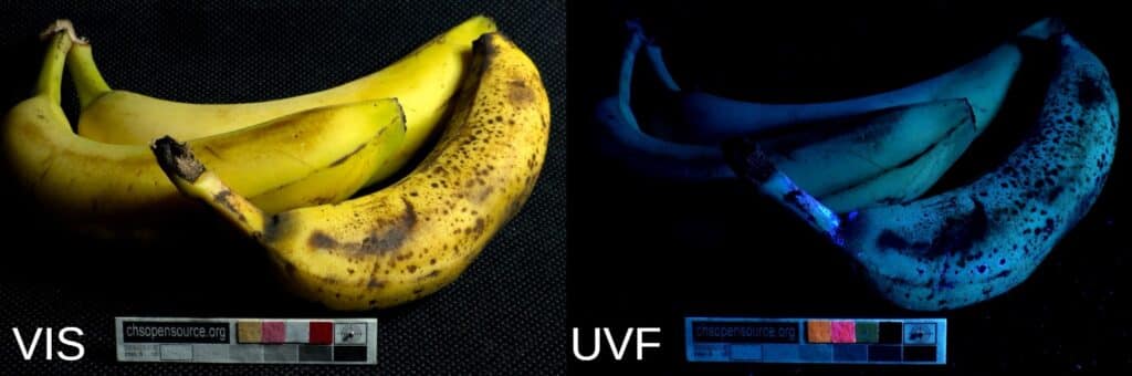
We start with bananas. Under UV illumination the peel shows a bluish fluorescence across its surface. When bananas become ripe—and especially when they are overripe—the fluorescence becomes much stronger. This bright blue glow is linked to the degradation of chlorophyll that occurs during the ripening process.
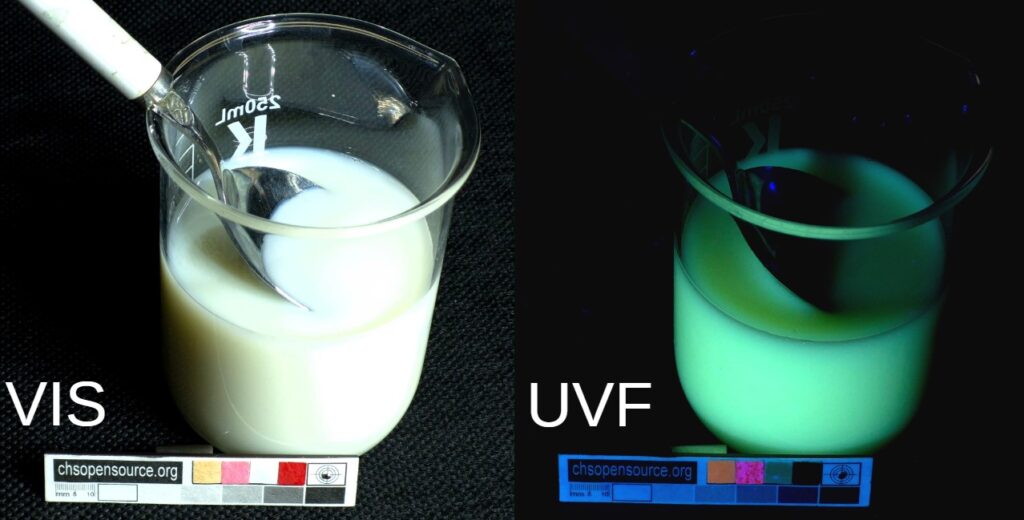
From the intense blue of bananas we move to the pale yellow-green fluorescence of milk, which appears soft and subtle under ultraviolet light.
Milk shows pale yellow-green UV fluorescence mainly due to riboflavin (vitamin B₂), a naturally occurring vitamin in milk that strongly fluoresces under ultraviolet light. Other components, such as certain proteins and aromatic amino acids, can contribute slightly, but riboflavin is the primary compound responsible for milk’s characteristic fluorescence.
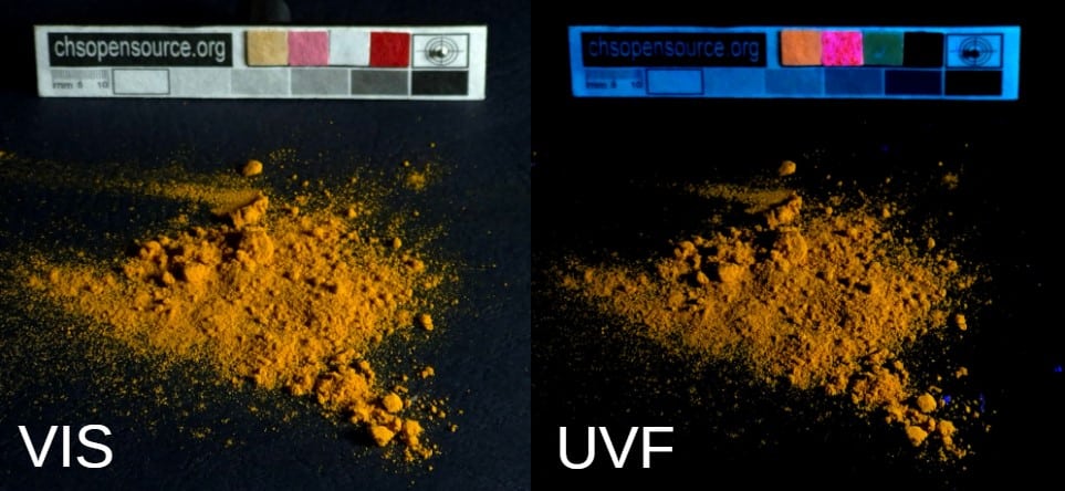
Next comes a dramatic shift in color with turmeric (curcuma). When illuminated with UV light, this common kitchen spice produces a very bright yellow fluorescence, revealing why compounds in turmeric have also been used historically as pigments and dyes.
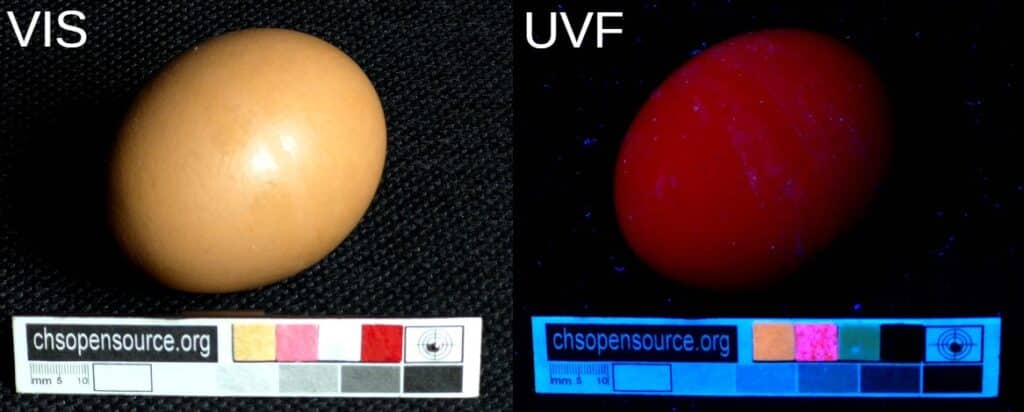
From bright yellow we move to the intense red fluorescence of eggshells. Under UV illumination the shell emits a vivid red glow caused by naturally occurring porphyrin compounds.
In many bird species, especially chickens, the pigment protoporphyrin IX is deposited on the eggshell during the final stage of egg formation in the shell gland (uterus) of the oviduct. This pigment gives brown eggs their characteristic color.
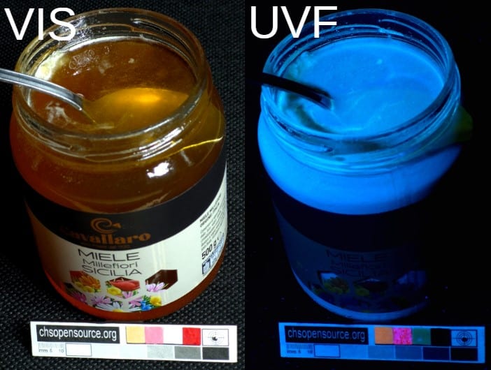
Finally, we finish with honey, which shows a soft whitish fluorescence with a slight bluish tone.
Honey shows weak whitish to bluish fluorescence under UV light because it contains several naturally fluorescent molecules.
The main contributors are phenolic compounds and flavonoids, which originate from the nectar of flowers collected by bees. These plant-derived molecules often fluoresce when excited by ultraviolet radiation.
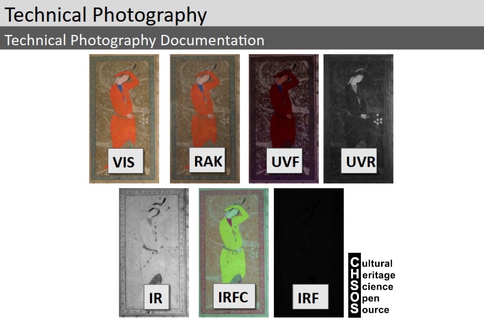
Technical Photography is one of the most powerful—and often overlooked—tools for the scientific examination of art and archaeology. If you are a conservator, scientist, or art collector and you are not yet familiar with this method, it is truly a missed opportunity. Using simple, affordable equipment and a clear methodology, Technical Photography allows you to reveal underdrawings, retouchings, material differences, and conservation issues in a completely non-invasive way. Far from being complex or inaccessible, it is an easy entry point into scientific analysis. In many cases, Technical Photography represents the first essential step toward a deeper understanding of artworks and archaeological objects.
Scientific Art Examination – Resources:
Getty Conservation Institute (GCI) – USA
The British Museum – Scientific Research Department – UK
Scientific Research Department – The Metropolitan Museum of Art, New York, USA
C2RMF (Centre de Recherche et de Restauration des Musées de France) – France
Rijksmuseum – Science Department – Netherlands