- Understand the origin of argon peaks in XRF spectra.
- Analyze the impact of various filter setups on the visibility of argon peaks.
- High-density polyethylene (HDPE) plate
- Bismuth cube
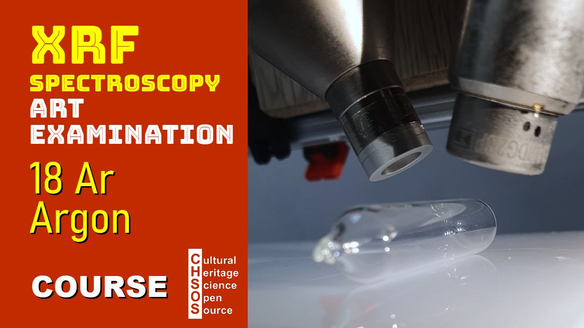
- Argon vial
- Plasma sphere
- Introduction to Argon in XRF Spectroscopy:
- Explain the presence of argon in the air and its interaction with X-rays.
- Discuss the characteristics of argon peaks (K alpha at 3 keV and K beta at 3.2 keV).
- Experiment 1: HDPE Plate Analysis:
- Use the HDPE plate to reflect X-rays and observe argon peaks under three filter setups: no filter, low keV filter, and routine filter.
- Compare spectra and identify argon peaks in each configuration.
- Experiment 2: Bismuth Cube Analysis:
- Analyze the bismuth cube with the same filter setups.
- Evaluate the intensity and visibility of argon peaks across configurations.
- Experiment 3: Argon Vial and Plasma Sphere:
- Attempt to detect argon directly using an argon vial under the spectrometer.
- Demonstrate argon excitation with a plasma sphere to confirm its presence visually.


Free Course: XRF Spectroscopy for Art Examination
The course XRF Spectroscopy for Art Examination introduces conservators, art historians, and scientists with interest in Art to the principles and practical applications of X-ray fluorescence (XRF) spectroscopy in the examination of artworks. The course starts with basic principles of XRF and gradually explores its role in identifying materials and methods used in the creation and conservation of art.
Course Objectives
- Understand the fundamentals of XRF spectroscopy and how it applies to the analysis of art.
- Learn the key features and limitations of XRF for examining art and archaeology.
- Gain skills in interpreting XRF spectra to identify specific elements in paint layers, inks and metals.
Scientific Art Examination – Resources:
Getty Conservation Institute (GCI) – USA
The British Museum – Scientific Research Department – UK
Scientific Research Department – The Metropolitan Museum of Art, New York, USA
C2RMF (Centre de Recherche et de Restauration des Musées de France) – France
Rijksmuseum – Science Department – Netherlands


