- Understand the presence and roles of calcium in painting preparation layers and pigments as a filler.
- Explore the application of XRF spectroscopy to detect calcium in different materials.
- Analyze historical changes in calcium use in paper manufacturing through spectra comparison.
- Pigments Checker: acrylic binder, chalk, bone black.
- Samples:
- Historical papers (16th, 18th, 19th century)
- Modern copy paper
- XRF spectrometer (no-filter setup)
- Discuss the prevalence of calcium in painting preparation layers.
- Examples: Gypsum-based layers (calcium sulfate) in Italian Renaissance paintings; calcium carbonate layers in Northern European art.
- Explain calcium as a filler in pigments and its implications for XRF analysis.
- Note that calcium signals often originate from the ground preparation or filler rather than the pigment itself.
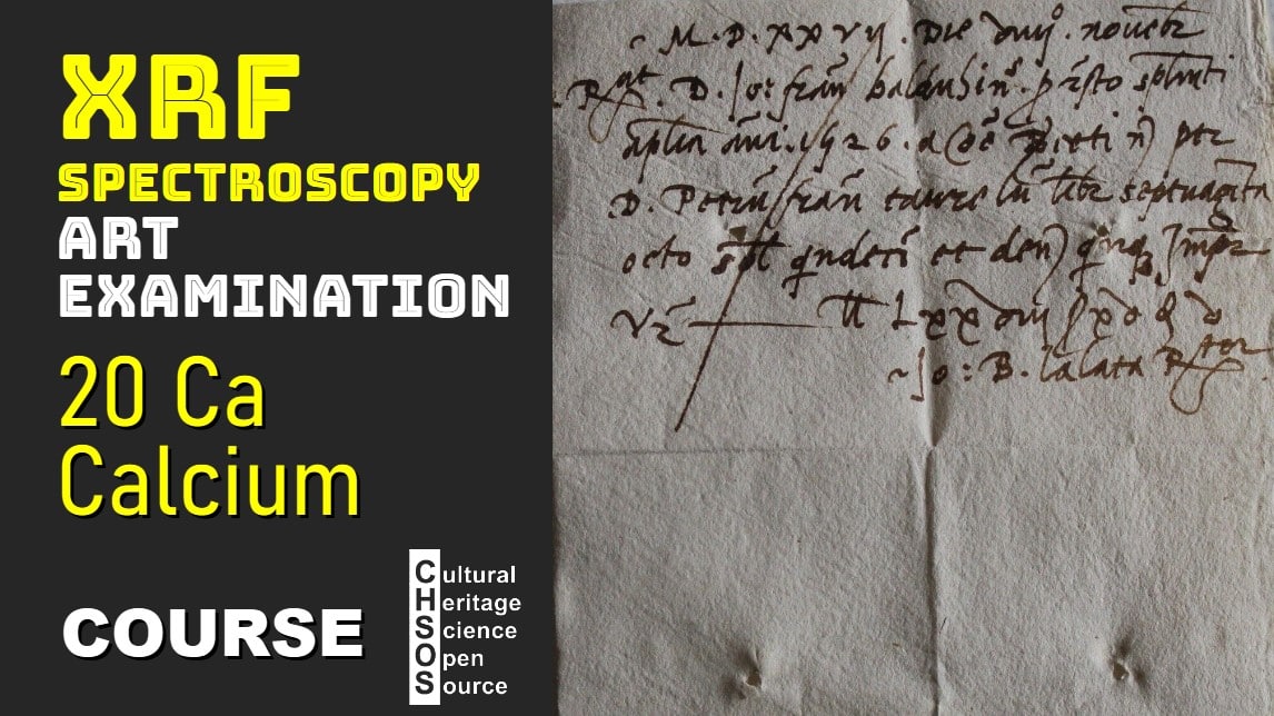
- Introduce calcium’s K-alpha (3.7 keV) and K-beta (4 keV) peaks, emphasizing their separation and detectability.
- Use the Pigments Checker to demonstrate calcium detection:
- Test a cardboard sample with an acrylic binder, highlighting strong calcium signals.
- Analyze chalk (calcium carbonate) and compare spectra, noting the prominent calcium peak.
- Examine bone black pigment and discuss its calcium phosphate content from charred bones.
- Analyze paper samples from different centuries using XRF spectroscopy:
- 16th-century paper (1527): High calcium peak (30,000 counts).
- 18th-century paper (1799): Reduced calcium content.
- 19th-century notary paper (1898): Minimal calcium signal.
- Modern copy paper: Substantially higher calcium content.
- Discuss how calcium levels reflect changes in paper manufacturing technology and practices over time.


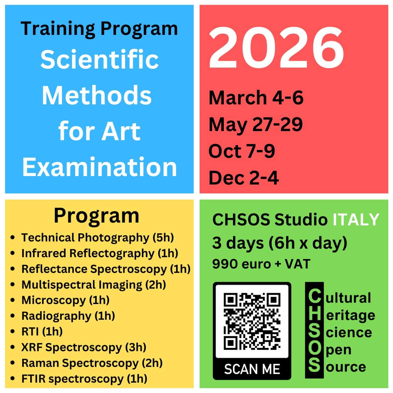
Free Course: XRF Spectroscopy for Art Examination

The course XRF Spectroscopy for Art Examination introduces conservators, art historians, and scientists with interest in Art to the principles and practical applications of X-ray fluorescence (XRF) spectroscopy in the examination of artworks. The course starts with basic principles of XRF and gradually explores its role in identifying materials and methods used in the creation and conservation of art.
Course Objectives
- Understand the fundamentals of XRF spectroscopy and how it applies to the analysis of art.
- Learn the key features and limitations of XRF for examining art and archaeology.
- Gain skills in interpreting XRF spectra to identify specific elements in paint layers, inks and metals.

Scientific Art Examination – Resources:
Getty Conservation Institute (GCI) – USA
The British Museum – Scientific Research Department – UK
Scientific Research Department – The Metropolitan Museum of Art, New York, USA
C2RMF (Centre de Recherche et de Restauration des Musées de France) – France
Rijksmuseum – Science Department – Netherlands