Objectives
- Understand the properties of magnesium relevant to XRF spectroscopy.
- Analyze the detectability of magnesium compared to sodium in handheld XRF systems.
- Explore the relationship between magnesium concentration and XRF peak visibility.
- Learn practical applications of magnesium detection in materials and pigments.
Materials
- Magnesium metal cube standard.
- Sample materials: pencil sharpener (magnesium alloy), magnesite (magnesium carbonate), celadonite, and green earth pigment.
- Online X-ray attenuation calculation tool (optional for demonstration).
Lesson Plan
- Recap of Previous Lesson
- Review sodium detection limitations using handheld XRF systems.
- Discuss the challenges posed by air attenuation of low-energy X-rays, particularly for sodium at 1 keV.
- Introduction to Magnesium Detection
- Highlight magnesium’s higher K-alpha line energy (1.25 keV) compared to sodium.
- Discuss the improved probability of magnesium X-rays reaching the detector.
- Theoretical Insights
- Use an online tool to calculate X-ray attenuation for magnesium.
- Compare the percentage of magnesium X-rays reaching the detector (10%) versus sodium (1%).
- Practical Tests and Results
- Test 1: Analyze a magnesium metal cube using the no-filter setup.
- Observe a distinct peak at 1.25 keV, confirming magnesium detection.
- Test 2: Analyze a pencil sharpener (magnesium alloy).
- Note similar results, with magnesium detected despite the presence of other alloying elements like aluminum.
- Test 3: Test magnesite (magnesium carbonate).
- Highlight reduced magnesium concentration and reduced peak intensity.
- Test 4: Test celadonite mineral.
- Discuss why magnesium detection fails due to low concentration in these samples.
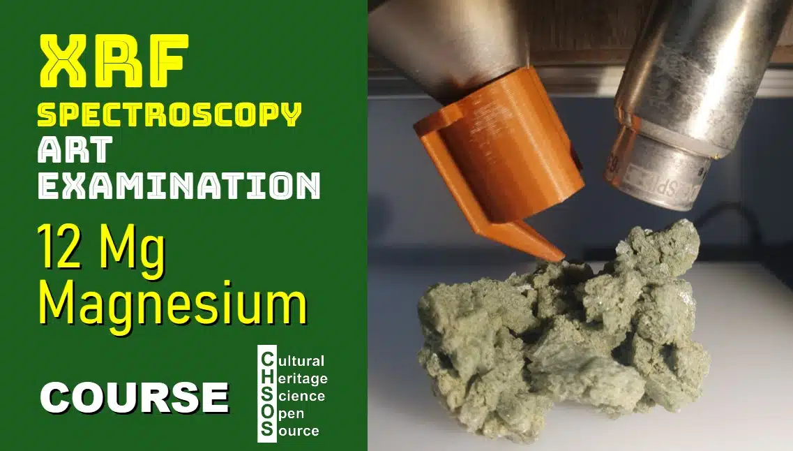
- Test 5: Examine green earth pigment using PIGMENTS CHECKER.
- Note the absence of magnesium detection despite its presence in the pigment composition.
- Test 1: Analyze a magnesium metal cube using the no-filter setup.
- Discussion and Interpretation
- Summarize findings: Magnesium is detectable in high-concentration forms (e.g., pure metal or alloys).
- Explain the challenges of detecting magnesium in low-concentration samples, especially in pigments and minerals.


Free Course: XRF Spectroscopy for Art Examination

The course XRF Spectroscopy for Art Examination introduces conservators, art historians, and scientists with interest in Art to the principles and practical applications of X-ray fluorescence (XRF) spectroscopy in the examination of artworks. The course starts with basic principles of XRF and gradually explores its role in identifying materials and methods used in the creation and conservation of art.
Course Objectives
- Understand the fundamentals of XRF spectroscopy and how it applies to the analysis of art.
- Learn the key features and limitations of XRF for examining art and archaeology.
- Gain skills in interpreting XRF spectra to identify specific elements in paint layers, inks and metals.

Scientific Art Examination – Resources:
Getty Conservation Institute (GCI) – USA
The British Museum – Scientific Research Department – UK
Scientific Research Department – The Metropolitan Museum of Art, New York, USA
C2RMF (Centre de Recherche et de Restauration des Musées de France) – France
Rijksmuseum – Science Department – Netherlands