Contributed by Samantha Stout
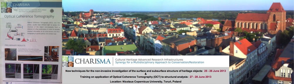
This post covers the second 2-day workshop held by CHARISMA partners in Torun, Poland from 27-28 June 2013.
This time the sessions were all geared towards OCT, a technique established for use in ophthalmology and then further developed by Prof. Targowski’s research group at UMK who’ve built their own system, which is particularly well adapted for use on cultural heritage objects. Attendees were first presented with a very detailed and informative introduction to the technique, where the different modalities (time domain, Td; sweep source, SS; spectral domain, SD; and Fourier domain, Fd) for capturing the tomographic signal were explained. (Additional information can be found on the, aptly titled OCT4ART webpage, where most papers are available in PDF format)

2. Torun Living Museum of Gingerbread. 3. Statue of Nicolaus Copernicus, Rynek Staromieski, (main square). 4. OCT Poster from CHARISMA Workshop.
OCT uses a low power, broadband light source, usually centered in the red or near-infrared region of the spectrum to allow for maximum transparency of painted layers. Because the light intensity is kept to a minimum, the technique is safe for use on the precious artifacts. OCT is particularly useful at visually presenting the layers beneath the surface of a given artifact in a cross-sectional form, and doing so non-invasively. That being said, the entire concept is based on the absorbance and scattering of light. So, once the source has been fully absorbed, there is no way to increase the information gained (with respect to what lies even further down). Additionally, in the tomogram we view interfaces specifically, and sensitivity is on the order of 1um; therefore, layers can be precisely measured and even mapped (and re-sliced) in the form of a 3D spectral cube constructed from data scanned over a region. It must be kept in mind that information on what the layers are made out of is not necessarily obtained. Usually conjectures about material content are made after examination with complementary techniques has been carried out.
Two talks, in particular, highlighted the unique potential that OCT brings to the table because it is especially suited towards identifying surface structure and layers. These presentations covered the concept of precisely and objectively monitoring laser cleaning and restoration procedures, and were given by Paraskevi Pouli of FORTH, in Greece, and Marcin Sylwestrzak of UMK. In my opinion, this is perhaps where OCT will be most utilized and will have the greatest impact on instituting new procedures with another level of verification to not do any harm when treating an artifact.
The hands-on portion of the training sessions was held on the last day for the majority of the afternoon, and included OCT examination demonstrated on a painting, a piece of stained glass, glazed ceramics, and fragments of jade artifacts. Specifically, the demonstrations were conducted to show both the research developed UMK system and a commercially available OCT system offered by ThorLabs GMBH. Both systems performed comparably and it was useful to see the technique in action.
OCT, in its currently developed form, is most useful on paintings for visualizing multiple layers of varnish, areas presenting craquelure or surface damage, and in some cases, repainted sites or areas of previous restoration.

There is some tuning of the instrument required to acquire a representative and crisp tomogram. And, most notably, there is a bit of a hurdle of instruction and training required to accurately interpret the tomographic result. This occurs because every cultural artifact is certainly a unique scenario, and a moderately complex understanding of the principles of operation and the signal post-processing is required to obtain the useful minute details of the particular artifact’s surface and sub-surface structure. In fact, a previously developed intimate knowledge of the artifact is most useful when attempting to interpret the tomogram, rendering this technique somewhat secondary with respect to diagnostic methods for culture heritage artifacts.
Through this workshop, which was completely free to attend, the CHARISMA partners have certainly opened the OCT technique to a wider audience. Unique, and praised for its non-invasive nature, OCT has potential to provide CH users with details previously not accessible without employing a micro-destructive technique.